A meningioma is a tumor that grows from the lining around the brain. Most are benign (not cancer), but as they grow they can press on the brain and nerves.
The patient’s story
A 56-year-old patient had repeated headaches, changes in behavior and problems with vision in both eyes.
Diagnosis
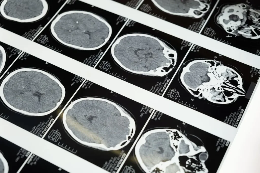
A brain MRI showed a large tumor at the base of the skull behind the eyes (the planum sphenoidale), pressing on the frontal lobes of the brain. The appearance was typical of a meningioma.
Treatment with advanced technology
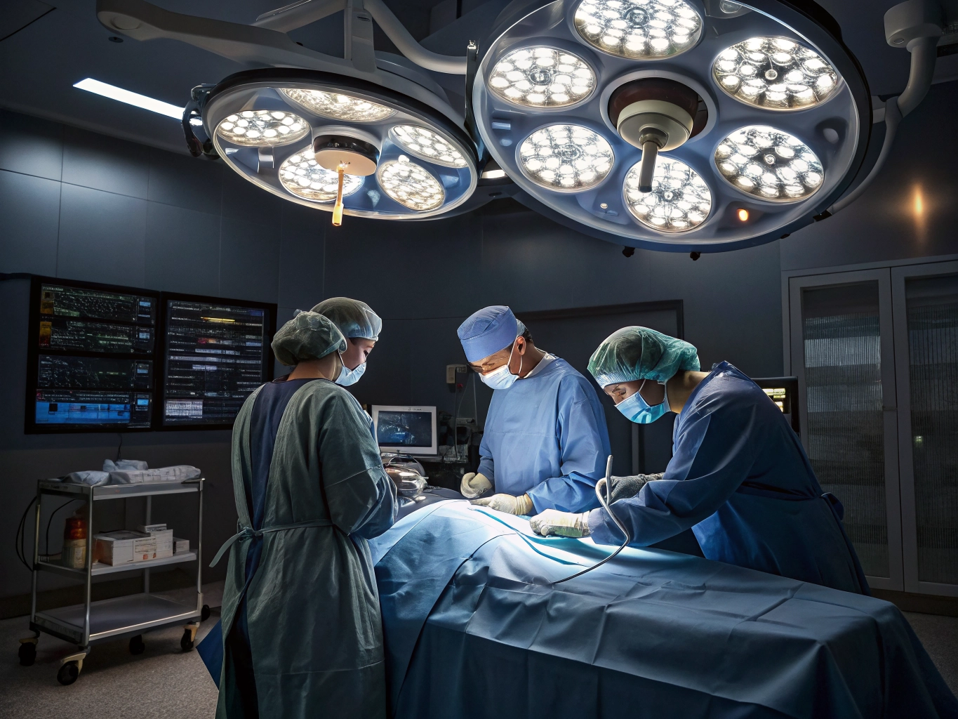
Our neurosurgeons removed the entire tumor through an opening at the side of the skull (pterional craniotomy). They used the KARL STORZ VITOM 3D exoscope, which gives a magnified, high-definition 3D view on a large screen. This allowed very precise work around the nerves of sight and the major blood vessels. Ethio Tebib is currently the only center in the country offering this platform.
Outcome
The tumor was confirmed as a benign grade I meningioma, and scans showed no tumor left behind. The patient’s headaches improved markedly, neurological function was preserved and the patient returned to normal daily life.
What you should know
- New or worsening headaches with vision changes or personality changes should be checked with a brain scan.
- Most meningiomas are benign and can be removed successfully.
Related care at Ethio Tebib
Learn more about our Neurology & Neurosurgery Department and our Radiology Department in Addis Ababa.
The team behind this case
This case was managed by Dr. Selehadin Bediru, Neurosurgeon, and our Neurosurgery team and the dedicated team at Ethio Tebib Hospital.
Need advice?
To book an appointment, book online or call our short code 9000. We are on the road to Kolfe, Sefere Selam, Addis Ababa.
This case was first shared on Hakim. It is published with the patient’s identity protected. It is for general information only and is not a substitute for medical advice.