When a clinician recommends advanced imaging, the question often lands immediately: “Do I need an MRI or a CT scan?” Both tests can be life-changing tools, yet they work in different ways, answer different clinical questions, and feel different as a patient.
Choosing well is not about picking the “better” scan. It is about selecting the right tool for the body part, the symptom, the urgency, and your personal safety factors.
What each scanner is actually doing
A CT scan (computed tomography) is a sophisticated form of X-ray. The machine rotates an X-ray beam around you while detectors measure how much radiation is absorbed by different tissues. A computer reconstructs those measurements into cross-sectional “slices,” which can be stacked into detailed 3D views.
MRI (magnetic resonance imaging) takes a different approach. It uses a strong magnetic field and radiofrequency pulses to stimulate hydrogen atoms in the body. As those atoms return to their baseline state, they emit signals that are captured and converted into images.
That difference matters, because it shapes what each test is best at seeing and what risks must be considered.
The practical difference you notice first: speed vs. detail
CT is built for speed. In urgent care, emergency medicine, and trauma settings, that speed can be the whole point. Many CT scans capture the key images in seconds, with the full visit often finishing within minutes once preparation is complete.
MRI is built for contrast and characterization. It can take longer because it collects multiple “sequences,” each highlighting different tissue properties. The longer scan time is often the price paid for exceptionally rich soft-tissue detail.
A single sentence that is worth remembering: CT tends to answer urgent “what is happening right now?” questions quickly, while MRI tends to answer “what exactly is it?” questions with greater tissue contrast.
What MRI tends to show best
MRI shines when the problem involves soft tissues where subtle differences matter. The images can separate tissue types with striking clarity, which is why MRI is widely used in neurology and musculoskeletal medicine.
Common areas where MRI is often favored include the brain and spinal cord, discs and nerve roots, ligaments and tendons, cartilage and menisci, and many pelvic organs.
MRI can also be useful when clinicians need to map the extent of a lesion, evaluate inflammatory change, or monitor therapy response in a way that depends on tissue composition, not only anatomy.
What CT tends to show best
CT is excellent for bone detail and for air-filled regions. It is also very strong at finding acute bleeding and visualizing lungs with high clarity. When paired with iodinated contrast, CT angiography can rapidly assess blood vessels in the brain, chest, abdomen, and limbs.
CT is commonly selected for complex fractures, sinus disease, many chest concerns, kidney stones, suspected internal injury after trauma, and several causes of acute abdominal pain.
In many emergency protocols, CT comes first because it is fast, widely available, and provides reliable answers under time pressure.
Side-by-side comparison
| Category | MRI | CT |
|---|---|---|
| Core technology | Magnetic field + radiofrequency signals | Rotating X-ray beam + detectors |
| Radiation | None (non-ionizing) | Yes (ionizing radiation) |
| Strengths | Soft-tissue contrast, brain/spine, joints, many pelvic assessments | Speed, bone detail, lungs, acute bleeding, many urgent evaluations |
| Typical scan feel | Longer, loud, narrow bore; staying still matters | Shorter, quieter, wider opening |
| Common contrast agent | Gadolinium-based (when needed) | Iodinated (when needed) |
| Key limitations | Metal safety rules; claustrophobia for some; longer time | Radiation exposure; iodine contrast concerns in some patients |
Safety: what “safe” means for each test
Both tests are considered safe when ordered appropriately and performed with proper screening. The safety conversation is simply different.
CT safety is mostly about radiation dose. A single CT scan usually carries a low risk, yet repeated scans can increase cumulative exposure over time. This is one reason clinicians are careful with CT use in children and in patients who may require many follow-up exams.
MRI safety is mostly about magnets. The strong magnetic field can interfere with certain implants or attract ferromagnetic fragments. Screening is rigorous for pacemakers, cochlear implants, aneurysm clips, retained metal fragments, and other devices. MRI also requires hearing protection because the scanner can be very loud.
Pregnancy adds another layer. MRI without contrast is commonly considered acceptable in pregnancy when clinically needed, while CT of the abdomen or pelvis is generally avoided unless the medical need is pressing and alternatives are not suitable.
Contrast dye: not everyone needs it, but everyone should ask about it
Contrast can make crucial findings visible, yet it is not automatically required. The decision depends on the clinical question.
Gadolinium contrast (MRI) is generally well tolerated, with rare allergic reactions. The most important caution is in severe kidney impairment, where certain gadolinium agents may be avoided.
Iodinated contrast (CT) can also be well tolerated, though allergic-type reactions can occur. Kidney function matters here too, especially for people with known chronic kidney disease, dehydration, or other risk factors.
A helpful mindset is to treat contrast as a medication: it has benefits, it has risks, and it should be matched to the question being asked.
After you have discussed your history, these are smart items to bring up with the ordering team:
- Prior contrast reaction: hives, wheezing, swelling, or severe nausea after a previous scan
- Kidney disease: any known reduced kidney function, dialysis, or recent abnormal creatinine results
- Medication list: include metformin, anticoagulants, and any drugs your clinician flagged for procedures
The patient experience: what it feels like inside the room
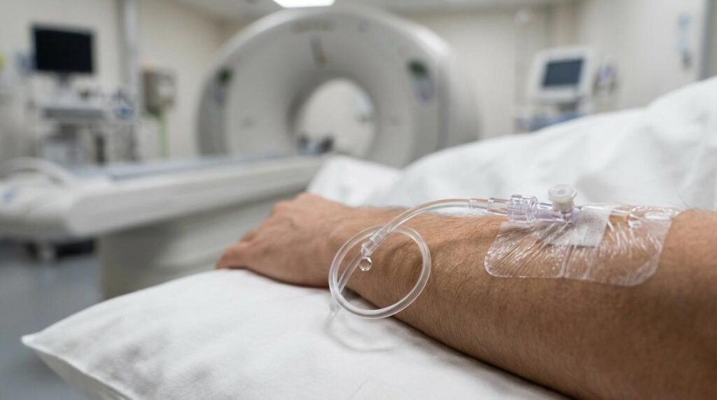
CT usually feels straightforward. You lie on a table that moves through a ring-shaped opening. You may be asked to hold your breath briefly. If contrast is used, you might feel a warm flush or metallic taste for a short time.
MRI can feel more intense simply because it lasts longer and is louder. Ear protection is standard. Some exams require breath-holds, and motion matters because the scanner collects many sequences over time.
If claustrophobia is a concern, tell the team early. Many imaging services can offer coaching, positioning adjustments, and in some cases mild sedation when appropriate.
How clinicians decide which test you might need
The decision is usually driven by three factors: what body system is involved, how urgent the situation is, and what diagnosis needs to be confirmed or ruled out quickly.
Here are common patterns clinicians use in real practice:
- Emergency speed: CT is often preferred when minutes matter, including major trauma or suspected acute bleeding
- Soft-tissue precision: MRI is often preferred for nerves, spinal cord, ligament injuries, and detailed brain assessment
- Lungs and bone detail: CT is often preferred for lung evaluation and complex fractures
Special situations: children, pregnancy, and chronic conditions
Children are more sensitive to radiation and have more years ahead for any radiation effects to matter. When MRI can answer the same question, clinicians often prefer MRI for pediatric patients, while also balancing the practical challenge that some children may need sedation to remain still for an MRI.
Pregnancy requires extra care. Ultrasound is often the first step for many pregnancy-related concerns. MRI without contrast may be used when needed. CT is generally reserved for situations where it is clearly justified, with careful technique to minimize exposure.
Chronic conditions often bring repeat imaging. In those settings, the lack of ionizing radiation can make MRI attractive when it provides the needed information, while CT remains valuable when speed, lung detail, or bone assessment is central to care.
Getting ready: what to do before you arrive
Preparation is usually simple, though it changes when contrast is planned. You may be asked to fast for a few hours before a contrast-enhanced study, and kidney function testing may be requested based on your risk profile.
MRI preparation focuses on metal safety. Expect to remove jewelry, watches, and other metal items and to answer detailed screening questions about implants or prior surgeries.
CT preparation focuses on contrast safety and pregnancy screening when relevant. Hydration can be encouraged when iodinated contrast is used, based on clinical guidance.
Access to both options under one roof
Many patients benefit when CT and MRI are available in the same hospital, because it supports fast escalation from one test to another when findings require clarification. It also helps when the care team needs imaging that fits a specific clinical pathway, whether that is emergency triage, surgical planning, oncology staging, or follow-up.
At Ethio Tebib Hospital, radiology services include both MRI and CT capabilities, with MRI performed on a 1.5 Tesla system and CT offered through modern multidetector scanners. The department also supports advanced protocols including CT angiography, along with digital image management through PACS to help clinicians review studies efficiently across services.
That mix matters most when care is time-sensitive, or when multiple specialists need to review the same images to plan treatment.
Results and next steps: what happens after the scan
Images are interpreted by trained radiology physicians, then shared with the referring clinician who integrates the report with your symptoms, exam findings, and laboratory results. Some findings lead to reassurance and routine follow-up. Others trigger a focused next step, like a specialty referral, a targeted lab test, or a procedure guided by imaging.
As the podiatry team at GetMyStride points out in their explainer on how to tell the difference between toenail fungus and a bruised or injured nail, careful history and examination can sometimes resolve the diagnosis without further imaging.
If you want to be an active partner in the process, ask one practical question: “What decision will this scan change?” It keeps the choice between MRI and CT grounded in clinical value, which is where the best imaging decisions are made.